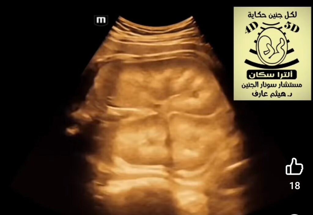
Fetal ultrasound for Auto somal Recessive Polycystic Kidney Disease (ARPKD) typically reveals symmetrically enlarged, highly echogenic (bright) kidneys, often detectable by the second trimester (around 18-24 weeks). The kidneys frequently show a “salt-and-pepper” or heterogeneous texture due to tiny cysts, leading to significantly reduced amniotic fluid (oligohydramnios). Ultrasound Findings (ARPKD):Renomegaly: Significantly enlarged kidneys (nephromegaly) that may fill the abdomen.Echogenicity: Hyperechoic (bright) kidney parenchyma, often without clear separation between the cortex and medulla.Cysts: While the disease is cystic, the cysts are often microscopic or too small to resolve (typically cm), differentiating it from the larger cysts in Autosomal Dominant PKD (ADPKD).Oligohydramnios: Decreased amniotic fluid, which is a significant indicator, often leading to pulmonary hypoplasia.Reversed corticomedullary differentiation: The medulla may appear brighter than the cortex. Timing and Progression:Early Detection: While sometimes seen earlier, characteristic findings are usually prominent in the second half of pregnancy.Serial Scans: Serial ultrasounds are used to monitor kidney enlargement and amniotic fluid appearance of ARPKD can overlap with other conditions. Similar findings include: Autosomal Dominant Polycystic Kidney Disease (ADPKD).Meckel-Gruber syndrome.Congenital nephrotic syndrome

 cm), differentiating it from the larger cysts in Autosomal Dominant PKD (ADPKD).Oligohydramnios: Decreased amniotic fluid, which is a significant indicator, often leading to pulmonary hypoplasia.Reversed corticomedullary differentiation: The medulla may appear brighter than the cortex. Timing and Progression: