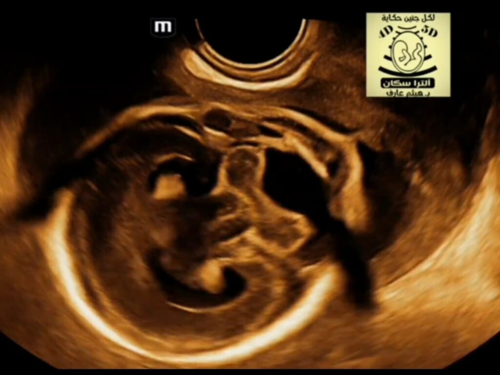
Fetal holoprosencephaly ultrasound
What is Holoprosencephaly (HPE)?Brain Malformation:HPE is a condition where the forebrain (prosencephalon) fails to divide completely during fetal development. Severity:HPE is classified into different types based on the degree of brain separation: alobar (most severe, no separation), semilobar (partial separation), and lobar (least severe, some separation). Ultrasound FindingsEarly Diagnosis: HPE can be diagnosed as early as the first trimester with ultrasound. Key Features:Absence of Septum Pellucidum: The thin wall separating the lateral ventricles is missing. Fused Ventricles: A single, fused ventricular cavity is present. Fused Frontal Horns: The anterior horns of the lateral ventricles appear fused and often have a squared-off appearance. Simplified Brain Structures: The interhemispheric fissure and falx cerebri (the dural fold that divides the brain) may be incompletely formed or absent. “Snake Under the Skull” Sign: In lobar HPE, a sagittal view with color Doppler can show the anterior cerebral artery located on the surface of the brain rather than in the midline fissure. Dorsal Cyst: A fluid-filled cyst may be present at the back of the head. Associated FindingsFacial Abnormalities: HPE is often associated with midface abnormalities, such as a single nostril (cyclopia) or cleft lip and palate. Polyhydramnios: An excess of amniotic fluid can occur due to impaired fetal swallowing.