pulmonary 🫁 sequestration fetal ultrasound
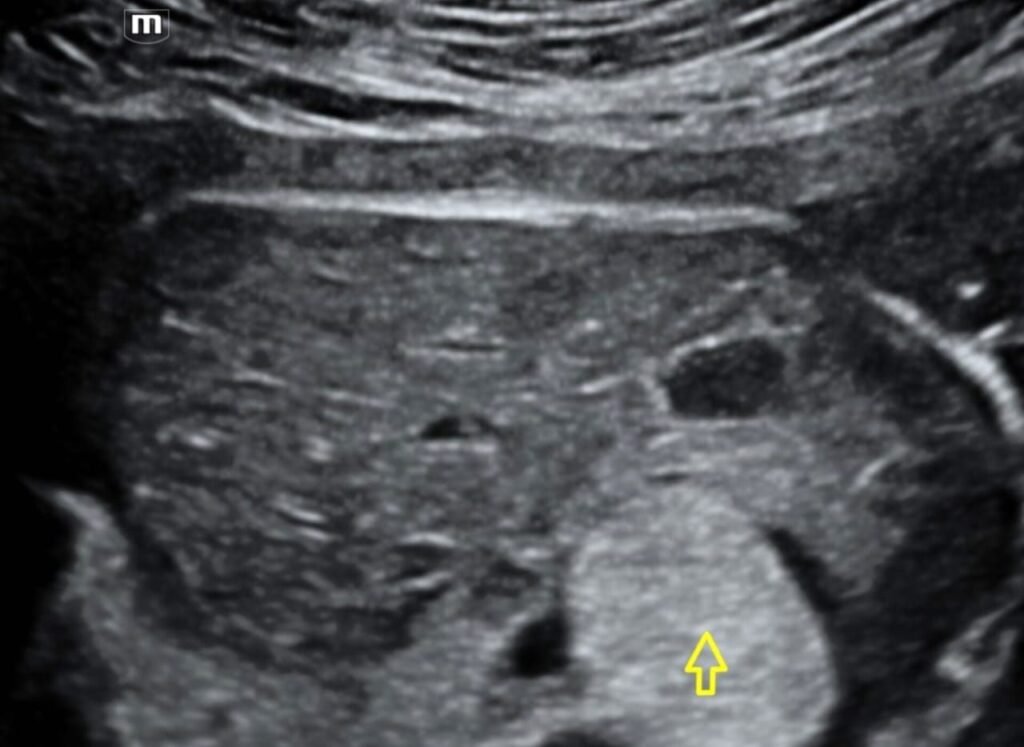
sequestration, also called accessory lung, refers to the aberrant formation of segmental lung tissue that has no connection with the bronchial tree or pulmonary arteries. It is a bronchopulmonary foregut malformation (BPFM).There are two types:intralobar sequestration (ILS)https://youtu.be/DIiLLGkgdR4?si=gvIsPHF5sufJZ2sHextralobar sequestration (ELS)https://youtu.be/QYFFFd5ntBg?si=vwElTw58a4bk6C89extralobar intrathoracicextralobar subdiaphragmaticClinical presentationExtralobar sequestration more commonly presents in newborns as respiratory distress, cyanosis, or infection ref. Intralobar sequestration presents in late childhood or adolescence with recurrent pulmonary infections ref, and in adults with cough, chest/back pain, dyspnea, and fever 16.PathologyPulmonary sequestration can be divided into two distinct groups based on the relationship of the aberrant segmental lung tissue to the pleura:intralobar sequestrationaccounts for the majority (75-85% of all sequestrations 4,5,7)present later in childhood with recurrent infectionsextralobar sequestrationless common (15-25% of all sequestrations 4,5,7)usually present in the neonatal period with respiratory distress, cyanosis, or infectionrecognized male predilection (M:F ratio ~4:1)can be infradiaphragmatic in ~10% of casesThe two types of sequestration are similar in their relationship to the bronchial tree and arterial supply/venous drainage but differ in their relationship to the pleura.By definition, there is no communication with the tracheobronchial tree. In the vast majority of cases, the abnormal lung tissue has a systemic arterial supply which is usually a branch of the aorta:intralobar sequestrationsvenous drainage commonly occurs via the pulmonary veins but can occur through the azygos-hemiazygos system, portal vein, right atrium or inferior vena cavaclosely connected to the adjacent normal lung and do not have a separate pleuraextralobar sequestrationsvenous drainage most commonly through the systemic veins into the right atrium (but is variable)separated from any surrounding lung by its own pleuraLocationOverall, sequestration preferentially affects the lower lobes. 60% of intralobar sequestrations affect the left lower lobe, and 40% the right lower lobe. Extralobar sequestrations almost always affect the left lower lobe, however, ~10% of extralobar sequestrations can be subdiaphragmatic .UltrasoundThe sequestrated portion of the lung is usually more echogenic than the rest of the lung. On antenatal ultrasound, an extralobar sequestration may be seen as early as 16 weeks gestation and typically appears as a solid well-defined triangular echogenic mass . Color Doppler may identify a feeding vessel (in-utero cases) from the aorta. If the sequestration is subdiaphragmatic, it may appear as an echogenic intra-abdominal mass. هتلاقيه بالظبط في الحاله ديhttps://youtu.be/QYFFFd5ntBg?si=vwElTw58a4bk6C89