غير مصنف
esophageal pouch sign fetal ultrasound
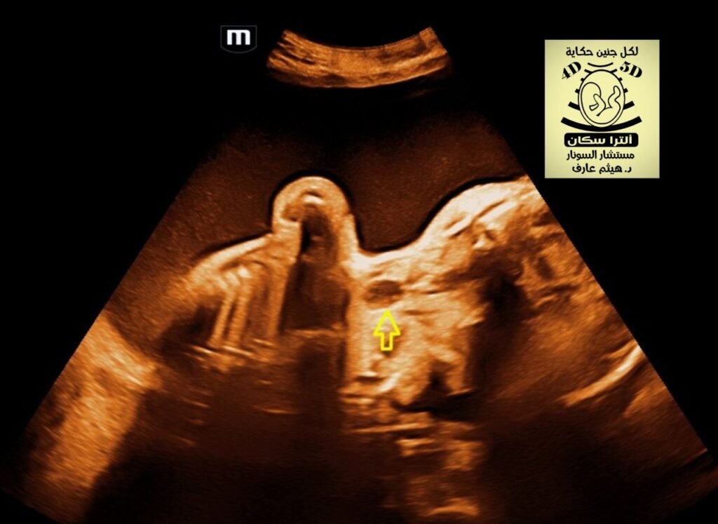
In fetal ultrasound, the
oesophageal pouch sign (or “upper neck pouch sign”) is a direct sonographic marker for esophageal atresia (EA). It is characterized by the visualization of a fluid-filled, blind-ending proximal esophagus that distends during fetal swallowing.
Imaging Features
- Appearance: A transient, anechoic (fluid-filled) cystic structure in the fetal neck or upper mediastinum.
- Dynamic Nature: The pouch typically enlarges as the fetus swallows amniotic fluid and then empties or disappears as the fluid is regurgitated.
- Timing: It is most commonly visualized after 26 weeks of gestation, though it has been reported as early as 22 weeks.
- Indirect Signs: It is often sought when indirect signs are present, such as polyhydramnios (excess amniotic fluid) and a small or absent fetal stomach bubble.
Clinical Significance
- High Specificity: While indirect signs like an absent stomach have low predictive value, the visualization of an esophageal pouch is highly specific for EA.
- Prognostic Value: The location of the pouch can help predict surgical outcomes.
- Mediastinal Pouch: Often associated with a shorter atretic gap and a better prognosis for primary surgical repair.
- Neck Pouch: Often indicates a longer gap between the proximal and distal esophagus, which may require more complex, staged surgeries.
- Tracheoesophageal Fistula (TEF): The sign can be seen regardless of whether a fistula is present.