
غير مصنف
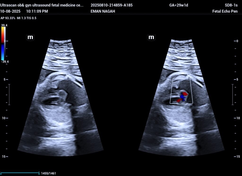
Pseudoaneurysm of mitral–aortic intervalvular fibrous area (MAIVF) fetal ultrasound
- Location:The P-MAIVF is situated between the mitral and aortic valves, specifically in the mitral-aortic intervalvular fibrosa (MAIVF).
- Formation:It’s not a true aneurysm, but rather a collection of blood outside the normal vascular space, contained by surrounding tissue.
- Causes:Commonly linked to infective endocarditis (bacterial infection of the heart valves) and surgical trauma to the aortic valve.
- Complications:Can lead to serious issues like fistulous tract formation (abnormal connection to other heart chambers), coronary artery compression, and even rupture.
- Symptoms:May include infection, chest pain, heart failure symptoms (shortness of breath), or neurological problems (like strokes).
- Congenital pseudoaneurysms of the mitral-aortic intervalvular fibrosa (P-MAIVF) are extremely rare,
is an avascular, fibrous structure that provides continuity between the anterior leaflet of the mitral valve and the aortic valve.
They can lead to:
Compression or obstruction of nearby structures.
Fistula formation (abnormal connections).
Blood clots within the pseudoaneurysm (thrombus formation).
Rupture, which can be life-threatening.
The exact cause of congenital P-MAIVF is not always clear, but it is believed to be related to underlying connective tissue abnormalities or developmental issues during heart formation.
ultrascan