Fetal right ventricular noncompaction cardiomyopathy (RVNCC)
🫀 CARDIAC FINDINGS: BY NOAH KIRUMIRA
Cardiomegaly with Right-Sided predominance: RV>LV, RA>LA, RV wall thickening ➡️ Suggests pressure/ volume overload of the Right heart.
🫀 RV shows rough, irregular internal wall contours and grooves compared to the LV – is highly suggestive of trabecular hypertrophy or Hyper-trabeculation, which raises the possibility of right ventricular non-compaction (RVNC) or a related cardiomyopathy.
💘 WHY IT MATTERS – In the normal fetal heart, the RV is more trabeculated than the LV (since the RV is the Dominant ventricle in Utero).
➡️ However, when the trabeculations are excessive, coarse, and associated with deep intertrabecular recesses, it goes beyond normal physiology and raises suspicion for non-compaction. In contrast, the LV is smoother and more compact is also an important comparative clue.
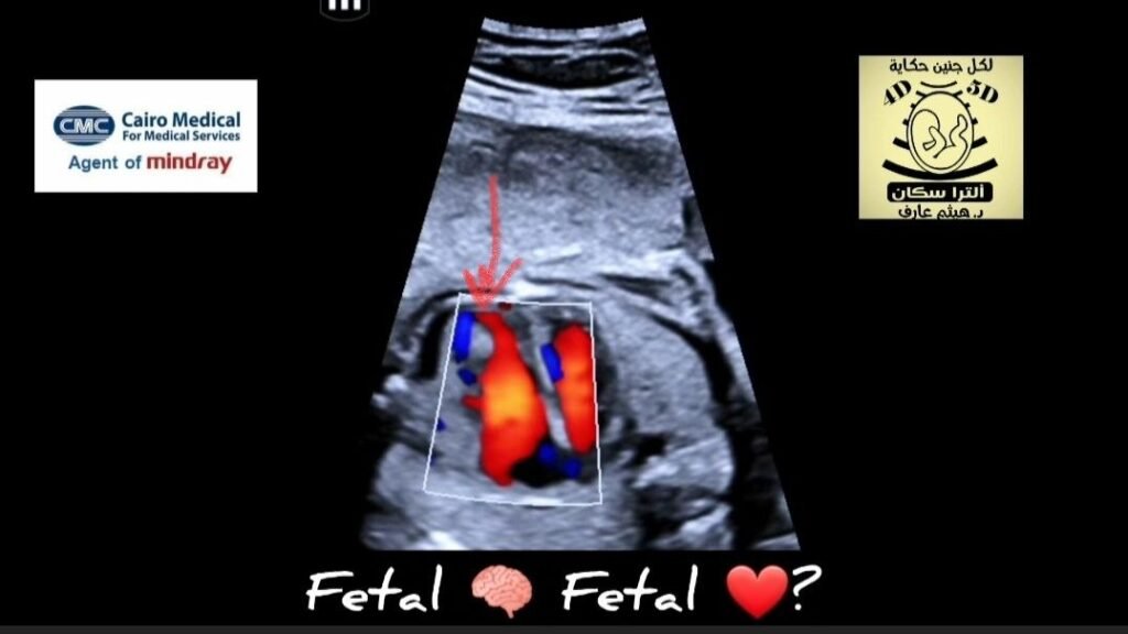
💗 Color Doppler demonstrates blood flow entering the deep recesses of a thickened, trabeculated RV wall, being one of the hallmark features of ventricular non-compaction Cardiomyopathy (NCCM) — Specifically RVNC.
🫀 Minimal pericardial effusion may be non-specific, but in the context of cardiomegaly, raises concerns for early cardiac decompensation.