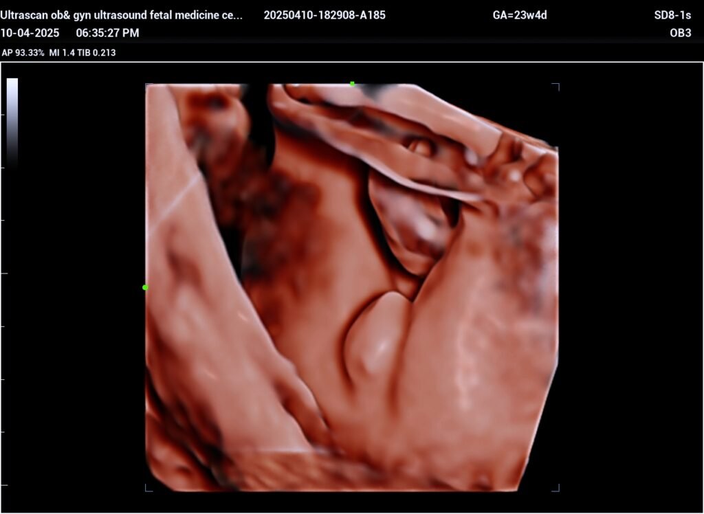
MICROPENIS FETAL ULTRASOUND ? Ambiguous genitalia
Prevalence:
1 in 5,000 births.
Ultrasound diagnosis:
Female fetus: clitoromegaly with normal labia.
Male fetus: micropenis, hypospadias, undescended testes, bifid scrotum.
On the basis of the cause, the condition is divided into:
True hermaphrodite: both ovarian and testicular tissue are found within the same gonad. The karyotype is female 46,XX, but there is a chromatinic material from the Y chromosome.
Female pseudohermaphrodite: virilized females with normal female karyotype and ovarian gonadal tissue. The causes include congenital adrenal hyperplasia (1 in 15,000), ingestion of androgens by the mother and maternal virilizing tumors.
Male pseudohermaphrodite: undervirilized males with normal male karyotype and testicular tissue. The causes include inadequate synthesis of testosterone or the presence of an androgen receptor defect.
Associated abnormalities:
Chromosomal abnormalities, mainly trisomy 13, triploidy and 13q syndrome, are found in a few cases.
The condition is commonly associated with genetic syndromes:
Smith-Lemli-Opitz syndrome: autosomal recessive; ambiguous genitalia, microcephaly, cardiac, renal and gastrointestinal defects, syndactyly and polydactyly.
WAGR syndrome: sporadic; Wilms tumor, aniridia (absence of the iris), geniturinary malformations, neurodevelopmental delay.
Other defects, mainly facial clefts and cardiac defects are often found.