غير مصنف
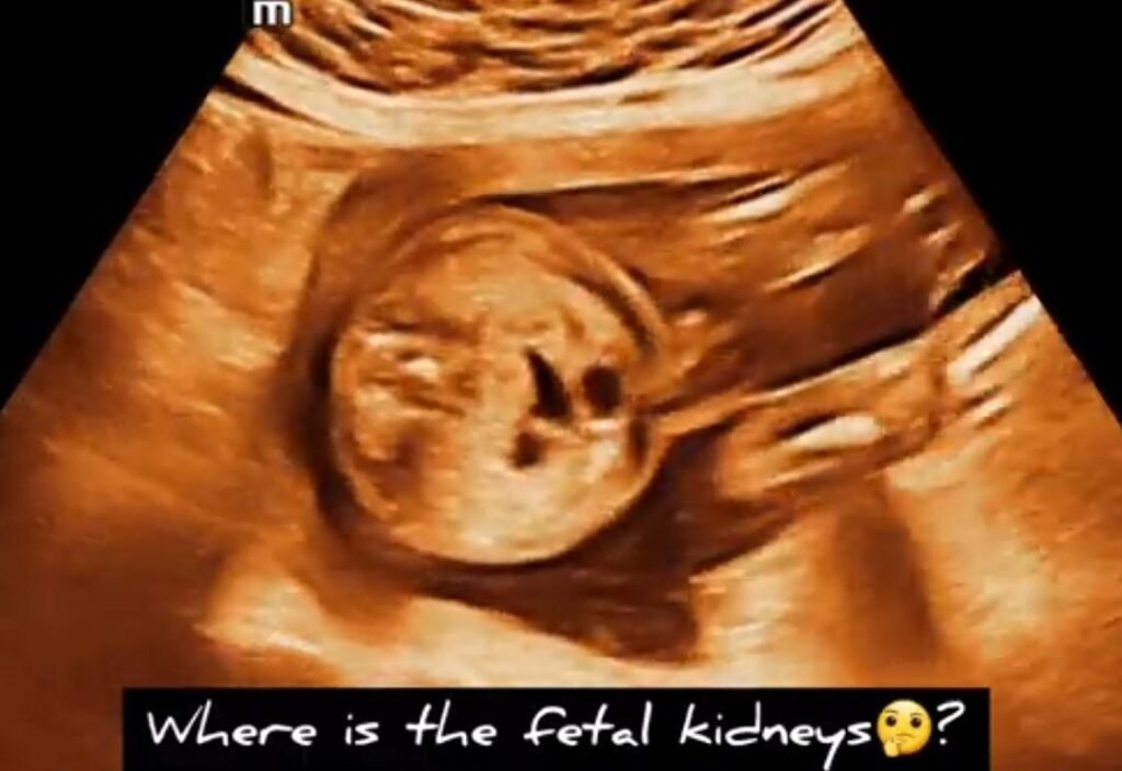
Pelvic kidney fetal ultrasound
Prevalence:
- 1 in 1,000 births.
Ultrasound diagnosis:
- Visualisation of the kidney in the pelvis above the bladder.
- Empty renal fossa on the affected side with the adrenal gland filling the space.
- Color Doppler might be useful to locate the hilum of the ectopic kidney.
- Normal bladder and amniotic fluid volume.
Associated abnormalities:
- The incidence of chromosomal defects and genetic syndromes is not increased.
Investigations:
- Detailed ultrasound examination.
Follow up:
- Ultrasound scans every 4 weeks to detect possible late-onset hydronephrosis.
Delivery:
- Standard obstetric care and delivery.
Prognosis:
- Good prognosis. Vesico-ureteral reflux and uretero-pelvic junction obstruction may occur.
Recurrence:
- No increased risk of recurrence.