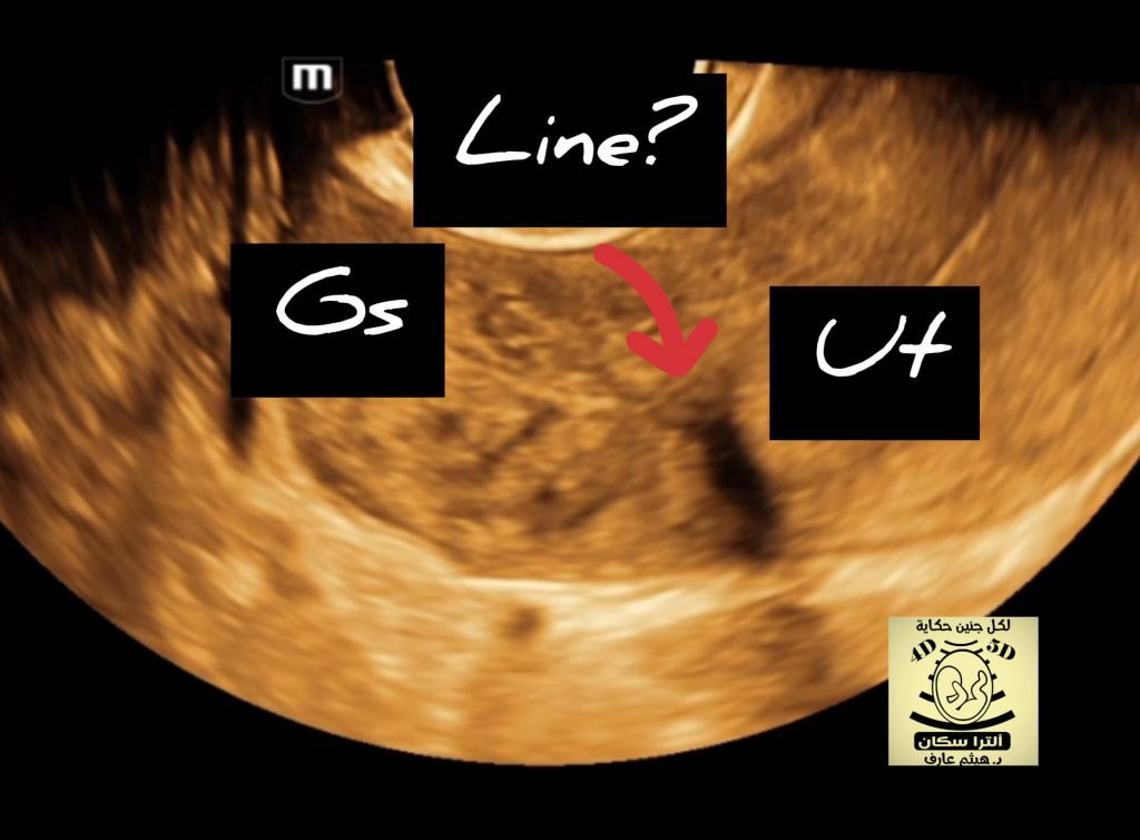
Cloacal Exstrophy (OEIS Syndrome)
collage pic by talented Dr ALAA OBEID
Overview
Cloacal exstrophy (OEIS Syndrome) is a rare and complicated condition that affects the lower abdominal wall structures of infants In utero, occurring in 1 out of 200,000 pregnancies and 1 in 400,000 live births. It is often diagnosed prenatally (before birth) by a fetal ultrasound and confirmed at the time of birth.
In infants born with this condition, the development of the lower abdominal wall (ventral) is interrupted during its normal embryonic migration and folding leaving many of the inner-abdominal structures exposed. These structures include the bladder, intestines and reproductive organs.
The pattern of inheritance for Cloacal exstrophy is unknown due to the small number of patients.
Symptoms
Four features of Cloacal exstrophy are frequently found together, and referred to as an OEIS Complex
Omphalocele
The omphalocele may be a large protrusion outside the cavity of the abdomen, involving several organs including the spleen, liver and intestine. It may also be small where only a portion of the intestine protrudes from the abdominal cavity.
Exstrophy of the bladder and rectum
Development of the bladder In utero is separated into two halves and open. The colon and rectum are also open with a segment of the rectum situated between the halves of the bladder and located on the surface of the abdomen.
Imperforate anus
A congenital birth defect, the anus of a new born has not been formed at birth (perforated) and the colon may connect to the urethra, bladder or vagina through a fistula (small tract).
Spinal defects
Infants with cloacal exstrophy often are born with further spinal complications including spina bifida.
